Add fluorescence microscopy images to data registry.#4939
Add fluorescence microscopy images to data registry.#4939emmanuelle merged 9 commits intoscikit-image:masterfrom
Conversation
|
Hello @mkcor! Thanks for updating this PR. We checked the lines you've touched for PEP 8 issues, and found:
Comment last updated at 2020-09-02 19:26:18 UTC |
|
@emmanuelle fyi, and tagging @jni for review |
|
I vote for "lily" and "kidney" as names, it's a good idea to shorten them. |
Sold! Done f70dbf9. Yes, this way they're also in line with the other datasets in scikit-image, which are named with one word: Thanks for weighing in, @GenevieveBuckley! |
|
A function must be added and we need to had an example in examples/data. For the gallery, a new page must be created for 3D dataset and we need to think how we can provide a preview -- we do not have the ability to interact with images yet. |
@sciunto tomorrow I want to try something out with |
You could also load the image into napari, switch it to 3D view and then take a screenshot (I had to split the color channels into separate layers to give them different colormaps). The kidney tissue is a pretty thin slice, so it doesn't look quite as impressive in 3D as other datasets might. Feel free to use these in the docs if they work for you.
|


|
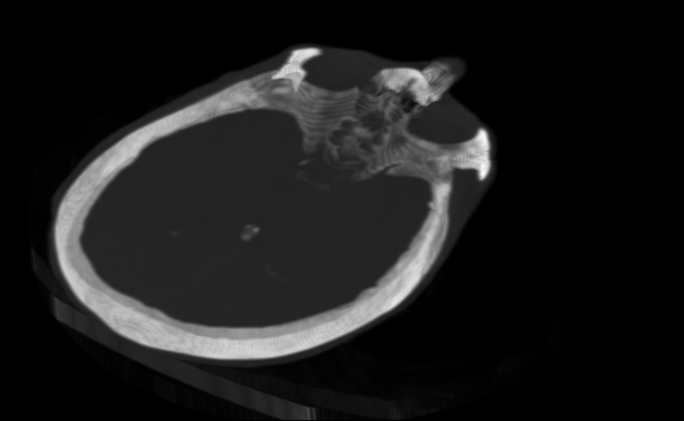
I don't know if you already have images in the docs for the brain image too, but it also looks pretty cool visualized in napari Axial plane 3D view |

|
It's a good idea, but we want to do not commit images to the main repository anymore. my taste for small incremental steps goes for the last solution. my 2 cents. |
|
I agree with @sciunto, we want to create our views programmatically from the data within our docs. Axis-aligned max projection is easy enough to do for now, and we can add something like the plotly Volume render eventually: https://plotly.com/python/3d-volume-plots/ (Though I don't really love those visuals...) |
|
@GenevieveBuckley and if you meant run napari within the docs, let's first figure that out in the napari docs! 😂 Also we don't yet have a camera model, though Nick has that as his next priority. |
Indeed, thanks for the pointer! |
@sciunto not sure I understand correctly: Why should the example(s) necessarily fall under https://scikit-image.org/docs/stable/auto_examples/#data? I was taking a stab at an 'application' example ("Longer examples and demonstrations"): #4946 |
@sciunto do you mean scikit-image/skimage/data/tests/test_data.py Lines 149 to 153 in 9e41809 |
Absolutely. The aim of this page is to display the content of our dataset |
Nope, I didn't mean that! We're in agreement here. I did not understand earlier that we wanted the figures/images for the docs to be auto-generated. |
I agree with this statement. The file you are looking for is here: |
|
@hmaarrfk yes, yes, spotted it! Thanks -- taking care of it now. |
|
|
||
| Returns | ||
| ------- | ||
| lily : (922, 922, 4) uint16 ndarray |
There was a problem hiding this comment.
Is this shape acceptable for our datasets?
I Feel like the output shape should be (4, 922, 922), in C-contiguous order
There was a problem hiding this comment.
4 is the number of colour channels: lily is a 2D multichannel image (https://scikit-image.org/docs/dev/auto_examples/applications/plot_3d_image_processing.html#explore-3d-images-of-cells). I'll add it in the docstring.
There was a problem hiding this comment.
Got it, yeah that wasn't clear to me. I was assuming x, y, z
skimage/data/__init__.py
Outdated
| def kidney(): | ||
| """Microscopy image of kidney tissue. | ||
| See `kidney-tissue-fluorescence.tif` entry at | ||
| https://gitlab.com/scikit-image/data/-/blob/master/README.md#data |
There was a problem hiding this comment.
I think there should be more fleshed out information here.
If you want to cross reference, then sphinx will have to fill things in for you.
I think it is important to be rather verbose in
https://scikit-image.org/docs/stable/api/skimage.data.html#skimage.data.brick
rather than sending the user around a multi click hunt.
|
Thanks @mkcor and reviewers! Could you just add |
Sure! Done 973508e. Good to know of this list. |
|
Merging, thanks ! We have some pretty bad CI failures :-(. But they are unrelated to this PR. |
Description
This PR is a simple follow-up on this one https://gitlab.com/scikit-image/data/-/merge_requests/6 by @GenevieveBuckley. We will then be able to load these images, that she obtained in confocal fluorescence microscopy, within scikit-image (via pooch).
We will thus have a 2D multichannel image (
lily-of-the-valley-fluorescence.tif) and a 3D multichannel image (kidney-tissue-fluorescence.tif) to play with! I'm excited for the upcoming gallery examples, notably more biomedical ones.The only hesitation I had when submitting this was whether I should keep the descriptive (short enough) filenames or shorten them into
lily.tifandkidney.tif, respectively.Let me refer to #3384 and #4601, and thank @GenevieveBuckley again!
Checklist
./doc/examples(new features only)./benchmarks, if your changes aren't covered by anexisting benchmark
For reviewers
later.
__init__.py.doc/release/release_dev.rst.